The biliary tract
The biliary tract is a part of the
The gallbladder
The gallbladder is a small, pear-shaped organ on the right side of the body, under
the right
The body can function without the gallbladder. If it needs to be removed because of disease or another problem, there are no serious long-term effects and the body can still digest food.
The bile ducts
The bile ducts are a series of thin tubes that connect the liver, gallbladder and duodenum to carry bile.
What the biliary tract does
The biliary tract stores, concentrates and transports
- bile salts
-
bile pigments (such as
bilirubin ) - fats (such as cholesterol)
- water
Bile drains from the liver through the intrahepatic bile ducts and flows into the hepatic duct. If the bile is not needed for digestion at that time, it flows into the cystic duct and then into the gallbladder, where it is stored. The gallbladder can store about 30 to 80 mL (6 to 16 tsp) of bile. The gallbladder absorbs water from the bile, making it more concentrated. When bile is needed for digestion after a meal, the gallbladder contracts and releases it into the cystic duct. The bile then flows into the common bile duct and into the duodenum where it will be used for digestion.
Structure of the gallbladder
The gallbladder is about 7 to 10 cm (3 to 4 inches) long and about 2.5 to 4 cm (1 to 1.5 inches) wide.
The gallbladder is made up of 4 four layers of tissue.
-
The mucosa is the inner layer of the gallbladder. It is made up of epithelial cells (epithelium) and loose connective tissue (lamina propria).
-
The muscular layer is a smooth muscle layer above the mucosa.
-
The perimuscular layer is a connective tissue layer that covers the muscular layer.
-
The serosa is the outermost layer that covers the outside of the gallbladder.
The gallbladder is connected to the liver and duodenum by a series of tubes (the bile ducts).
Structure of the bile ducts
The common bile duct is a very thin tube, about 10 to 12.5 cm (4 to 5 inches) long and less than 6 mm (less than 0.25 inches) in diameter.
A series of smaller ducts come together and drain into the common bile duct, which drains bile into the duodenum.
Many tiny tubes in the liver first collect bile from the liver cells. These tiny tubes join to form small ducts, which come together to form larger ducts called the right and left hepatic ducts. The right and left hepatic ducts exit the liver and then join together at the hilum (also called the hilar area) to form the common hepatic duct.
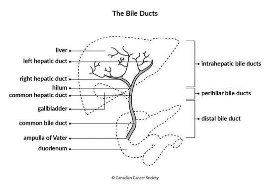
The common hepatic duct joins the cystic duct (which connects to the gallbladder) to form the common bile duct. The common bile duct passes through the pancreas before it empties bile into the duodenum. The lower part of the common bile duct drains into the duodenum through a channel called the ampulla of Vater.
The system of bile ducts is divided into 3 main segments based on their location: intrahepatic, perihilar and distal.
Intrahepatic bile ducts
The bile ducts inside the liver are called the intrahepatic bile ducts. The tiny tubes and small ducts that collect bile from the liver cells join together into larger and larger ducts, ending in the left and right hepatic ducts. These ducts drain bile from the right and left lobes of the liver.
Perihilar bile ducts
The first series of bile ducts outside the liver (called the extrahepatic bile ducts) are the perihilar bile ducts. They are located between the hilum and the point where the cystic duct and common hepatic duct join to form the common bile duct. Because the perihilar bile ducts are closest to the liver, they may also be called the proximal extrahepatic bile ducts.
Distal bile duct
The latter part of the extrahepatic bile ducts is the distal bile duct. It is farther away from the liver, located between the ampulla of Vater and the point where the cystic duct joins the common hepatic duct (but does not include these structures). The distal bile duct is made up of the common bile duct.